Home / About Us About Us
Interdepartmental Service Center of Veterinary Radiology was established on the basis of a joint project of the Departments of Veterinary Medicine and Animal Production and of Advanced Biomedical Sciences.

The Interdepartmental Center of Veterinary Radiology (CISRV) was established in 1985, to improve the use of resources and skills related to the development of morphological and functional Diagnostic Imaging with ionizing and non-ionizing radiation, within the veterinary disciplines and those related to them. As per the University Regulations, the Department of Veterinary Medicine and Animal Production (https://www.mvpa-unina.org) and from 26-10-04, the Department of Advanced Biomedical Sciences (http://scienzebiomedicheavanzate.dip.unina.it) join the CISRV of the University of Naples Federico II.
The CISRV takes care of the management and use of complex services and equipment commonly used by the Departments that proposed its establishment.
In addition to the service activity which translates into the radiographic, computed tomography and ultrasound examinations carried out daily, the CISRV promotes the teaching and research activities of the University Federico II of Naples, through scientific collaborations and the organization of a specialization course as well as courses and seminars.
Organization Chart
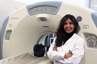
Administrative Secretary
Dr. Daniela Uccella
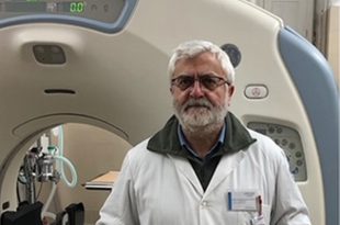
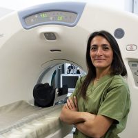
Professor
Leonardo Meomartino
Adelaide Greco
PhD Students
Dario Costanza
Erica Castiello
Fellows
Pierpaolo Coluccia
Equipment
The Interdepartmental Center of Veterinary Radiology has the following equipment:
- Eurocolumbus radiographic equipment, Program 80 US Eurostart, with anti-diffusion grid with Bucky-Potter system; this high-performance equipment (32 kW, max mA 1000, max kV 120, three-point parameter setting) allows you to perform direct and contrast radiographic studies of the thorax, abdomen, spine and skull, even on dogs of large and giant size, on small equids and small ruminants.
- • Agfa DR14e DR system with 35×43 cm plate, extremely fast (image reconstruction time < <2 sec.), with excellent contrast resolution and good spatial resolution (4.2 pairs of lines per mm).
- Agfa CR30X CR system with 6 cassettes (3 35×43 + 3 24×30), with good contrast and spatial resolution (5 pairs of lines per mm).
- 2 Workstations with Agfa Musica software (ver. 2 and 3) for management, viewing and archiving of RX exams.
- General Electric 16-layer spiral CT device mod. Brightspeed; this multilayer equipment allows to acquire studies of the whole body in less than a minute, even of large breed dogs, to obtain images with high spatial and contrast resolution, to carry out angio-urographic and dynamic studies.
- Philips Brilliance Workstation v. 4.5.5 for the management, display and archiving of CT exams, capable of performing multiplanar reconstructions, including curves, 3D, automated segmentations and volumetric and densitometric analyses.
- Esaote mod. Class C, with convex, microconvex and linear multifrequency probes, with modules for contrastographic studies and for the more superficial anatomical structures, capable of carrying out morphological, functional and Doppler studies, in small pets, exotics, and horses.
- Latest generation PACS system, based on open-source software (dcm4chee-arc-light version 5.11.1) for the management and archiving of images and video clips RX, TC and US in DICOM standard, with 24 TB capacity, redundancy of data through a RAID 6 configuration, possibility of consulting studies remotely via the company VPN network.
- Two Windows PC workstations for managing, viewing and archiving the RX, CT and US studies present in the Center’s PACS system.
- Mackbook Air M1 2020 PC for managing the patient database, the OVUD Center, and for reporting.
Other equipment:
- Hewlett Parker Expression A3 format transparency scanner mod. HP10000-XL for digitizing the Centre’s analogue film archive.
- Philips projector mod. UGO X-Lite for lessons, briefings, courses and seminars organized by the Centre.
- Gas anesthetic apparatus closed-circuit and open-circuit, with attached oxygen concentrator and waste gas aspirator for carrying out radiographic, CT and ultrasound studies under general anesthesia.
