Centro Interdipartimentale di Radiologia Veterinaria Università degli studi di Napoli Federico II
Home / Prestazioni / Direct radiographic and contrastographic examinations Direct RX Exam e Contrastographic
The services offered by our department are dedicated to all private and non-private users who need radiological investigations for animals
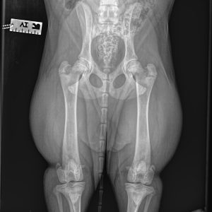
Radiography is the technique originally born together with the discovery of X-rays, which took place thanks to Roentgen in 1895. Radiography is considered the technique of choice for the skeleton and is currently used for the characterization of bone lesions, the effectiveness of treatments and monitoring of healing processes. In some cases, the radiographic examination can be performed on awake subjects, with the help of the owners, in the case of painful lesions or particular positions, it is necessary to subject the animal to deep sedation and/or general anesthesia.
Skeletal radiography is used to screen for many congenital or inherited bone and joint diseases, such as hip dysplasia and elbow dysplasia. In horses, with radiography it is possible to study the appendicular skeleton, the skull and the cervical spine, in the case of congenital, degenerative and traumatic lesions.
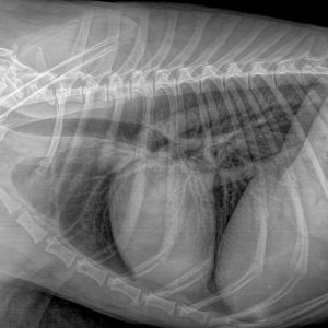
Radiography is also considered the technique of choice for studying the chest, especially the lungs. As with the skeleton, in cooperative subjects and with the help of the owners, the examination can be performed on an awake subject.
The radiographic examination of the abdomen is usually complementary to the ultrasound examination for the evaluation of some organs, in case of suspected foreign body and in the case of contrastographic examinations of the gastrointestinal tract.


